
MOVE4D: dynamic scanning technology for quantifying the efect of compression garments in runners
15 December 2025.
Autor/es: Alfredo Remón Gómez, José Peréz Maletzki*, Alfredo Ballester Fernández, Javier Gámez Payá, Sandra Alemany Mut.
Instituto de Biomecánica (IBV)
The Instituto de Biomecánica (IBV) has developed an innovative methodology to quantify soft-tissue displacement during sports practice. This methodology is based on MOVE4D dynamic scanning technology, which makes it possible to analyse, non-intrusively, variations in anthropometry and body shape in a subject in motion.
MOVE4D technology has been applied in an initial pilot study to assess the effect of compression garments in runners, enabling the anatomical modifications associated with their use to be identified and visualised. The results obtained show that this technology enables the objective measurement of soft-tissue displacement and demonstrate the ability of compression garments to reduce this displacement during running. MOVE4D is presented as a valid, non-intrusive technique for studying soft-tissue dynamics under real functional conditions. Its application opens up new opportunities to improve the design and development processes for compressive sportswear, facilitating the identification of the body areas where a higher level of compression needs to be applied and contributing to the definition of more appropriate compression levels for each body segment.
INTRODUCTION
Running is an activity that involves repetitive impacts that can reach up to 10 g, and ground reaction forces equivalent to between 1.5 and 3 times body weight in as little as 30 milliseconds [1]. All this causes vibrations in soft tissues (such as muscles, tendons, fat, ligaments and skin) and movements in different body systems. These vibrations can affect muscle function, the way energy is dissipated and, overall, running performance.
Soft-tissue vibrations can also cause neuromuscular fatigue, particularly in the legs. Several studies have shown that prolonged running alters muscle activation patterns, with notable increases in electrical activity (measured using electromyography, EMG) in muscles such as the medial gastrocnemius and vastus lateralis [2,3].
In addition, soft-tissue movement and vibrations are closely related to energy loss and running efficiency. Studies confirm that adjusting running technique to avoid unnecessary energy losses could improve an athlete’s performance [4].
Runners adapt their biomechanics to minimise the effects of soft-tissue vibrations and movement (STV). For example, changes in foot-strike pattern, stride frequency or pre-activation of muscles can reduce the amplitude and frequency of these vibrations. These adaptations are essential to maintain efficiency and reduce injury risk during prolonged runs [5–6].
Compression garments (CGs) are widely used in many sports to reduce and control soft-tissue vibrations and movement, and there is increasing interest in their design and in evaluating their performance. Various studies have found that, under certain conditions, compression garments can reduce blood lactate concentration during exercise [7]. In addition, their use has been reported to contribute to improved muscle oxygenation and faster re-oxygenation following exercise-induced fatigue [8–9]. The use of compression garments has also been linked to a reduction in impact force in activities such as vertical jumps. For example, custom-made compression shorts were observed to reduce impact force by 27% compared with non-compressive garments [10].
The most commonly used methodologies to analyse soft-tissue vibrations and movement are accelerometry, 3D motion analysis and surface electromyography (see Figure 1). However, all these methods share the same limitation: they can only perform localised measurements at specific points on the legs and are susceptible to errors caused by relative movement between the measurement system and the athlete’s equipment or skin [11].
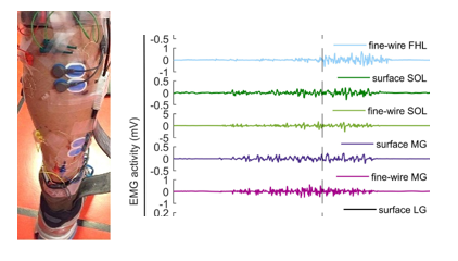
Figure 1. Example of measuring muscle activity using electromyography
In this context, it is necessary to move towards new methodologies based on 4D body-scanning technologies that make it possible to analyse soft-tissue movement throughout the entire running cycle in a global and direct manner. These approaches enable the simultaneous study of all relevant anatomical points on the body surface, reducing the limitations and potential artefacts associated with the use of external elements on the athlete, such as markers or sensors.
This article presents an innovative methodology based on dynamic 4D body scanning that makes it possible to quantify, objectively and non-intrusively, soft-tissue movement and shape changes during running, using dense three-dimensional (3D) information of the moving athlete’s body surface.
DEVELOPMENT
IBV has developed a study methodology based on MOVE4D dynamic body-scanning technology [12]. This technology makes it possible to capture the runner’s body shape with high accuracy at each instant of the running cycle. It is also a non-intrusive technique that does not interfere with the subject’s technique or movements, and records variations in body shape with millimetre precision and at high frequency.
The validation of the methodology was carried out using a pilot study that assessed soft-tissue movements in a group of runners. All subjects were regular runners (with a weekly training volume of at least 30 km/week) and had reported no injuries in the previous 3 months. The study subjects included runners with both rearfoot and midfoot strike patterns.
The compression garment evaluated in this study was a pair of medical compression stockings from the Jobst® brand (Stockholm, Sweden), designed to apply graduated compression, with a range from 22 (at the waist) to 29 mmHg (at the ankle). This progressive pressure profile seeks to optimise venous return and limit the accumulation of interstitial fluid.
The capture methodology consists of the following phases:
- An expert takes the runner’s body measurements and, using the manufacturer’s guide, selects the garment size to be used.
- 10-minute run on a treadmill as a warm-up. The last 5 minutes are performed under controlled running conditions. Speed and cadence are similar to the runner’s personal best half-marathon performance.
- Running capture with the Move4D system.
- Running capture with the Move4D system wearing the compression garment.
- Fatigue: the athlete runs outdoors for 1 hour.
- Fatigue level is assessed using the Borg 6–20 scale, with 17 being the value most commonly reported by the athletes.
- Running capture with the Move4D system.
- Running capture with the Move4D system wearing the compression garment.
Figure 2 shows an image of a capture session (left) and the Borg 6–20 scale used to assess fatigue level (right).
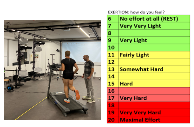
Figure 2. Image of a laboratory capture session (left) and Borg 6-20 scale for fatigue assessment
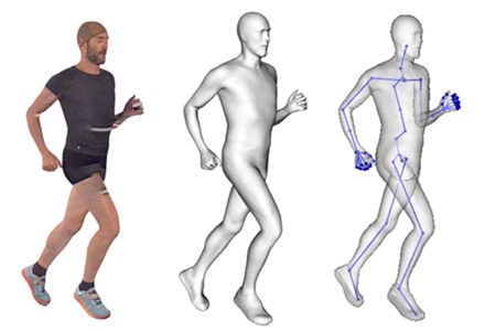
To ensure comparability of the different within-runner captures, it was verified that in all cases the athlete ran at the same speed and with the same stride cadence. Using the Move4D system, 3-second captures were recorded at a frequency of 178 fps, which ensured the capture of at least 3 complete strides. In total, four sequences were recorded for each athlete, each consisting of 534 frames (time instants). From these data, the MOVE4D system generated an avatar for each frame, accurately reconstructing the athlete’s body geometry in each of the 534 frames (see Figure 3) of the sequence.
Figure 3. Example of processing a frame: captured date (left), processed avatar (centre) and its skeleton (right)
Based on the information obtained using MOVE4D technology, different body metrics were calculated. The first morphological analysis performed was a study of variations in body-perimeter lengths throughout the sequence. In particular, six transverse contours were defined on each leg: three on the thigh and three on the calf. The location of these contours was established according to the length of each segment: the thigh contours were placed at 65%, 50% and 35% of the distance between the hip and the knee, while the calf contours were placed at 65%, 50% and 35% of the length of the knee–ankle segment. Figure 4 shows the six contours calculated by MOVE4D on a 3D avatar (left) and a graph describing the length of each contour throughout the sequence (right). In the graph, the four steps performed by the athlete in the captured sequence can be clearly identified through the periodicity of the signals.
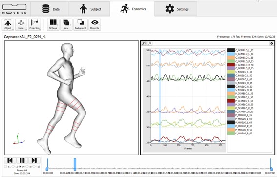
Figure 4. Representation on the avatar of the calculate body perimeters (left) and graph with perimeter values in the captured sequence (right)
Figure 5 shows a detailed analysis of variations in the perimeter length of the left calf measured at 35% of its length (knee–malleolus segment). Four biomechanically relevant instants of the running cycle are indicated on the graph using vertical dashed lines: the two foot strikes (heel strike) and the two mid-stance phases (midstance). Heel strike refers to the moment the foot contacts the ground, while midstance is the moment when the knees are in the same position in the sagittal plane. The graph shows the evolution of the perimeter across the four captures performed on the same runner. In order to enable reliable comparison between the time series, all captures were synchronised by aligning the start of each one with the first left-foot strike. Likewise, the horizontal dashed lines represent the perimeter with the user in a static standing position. The results show an approximate 10% reduction in perimeter associated with the fatigue state induced by continuous running, with this contraction observed both during running and in the static posture. This phenomenon may be attributed to loss of body mass—especially fluid components—derived from prolonged effort, as well as changes in muscle tension and redistribution of subcutaneous tissue. Additionally, use of the compression garment is associated with a slight additional reduction in contour length, consistent with the mechanical effect of external compression applied to the segment.
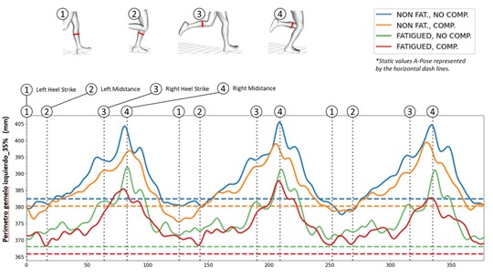
Figure 5. Study of the length of the left calf perimeter at 35% under the four condition studied
In a second phase of the analysis, the morphological variability of the calf perimeters throughout the running cycle was evaluated. To perform this analysis, the perimeters extracted from each frame of the captured sequence were superimposed on the same plane. The result is the superposition of between 300 and 400 contours comprising the full sequence of three strides from the athlete, with a darker tone observed in the most recurrent shapes and a lighter tone in the less frequent ones (Figure 6, left). From this representation, the perimeter shape variability index—Shape Variation Index (SVI)—is calculated, defined as the minimum area that covers all contour lines (Figure 6, right). This index quantifies the morphological variability of the perimeter and, therefore, the amount of soft tissue displaced in this area of the leg. A high SVI indicates greater contour instability, associated with more pronounced displacement of subcutaneous and muscle tissue.
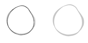
Figure 6. Aligned perimeters from the sequence (left). Area enclosed by the perimeters, or SVI (right)
Figure 7 shows the SVI calculated for the left calf at 35% of its length under the four experimental conditions of the study. As can be seen in the figure, in captures with compression garments, the calves tend to have a more rounded shape. Quantitatively, the SVI increases in the fatigued state, from which greater soft-tissue displacement under fatigue is inferred. Conversely, the use of compression garments is associated with a decrease in SVI.
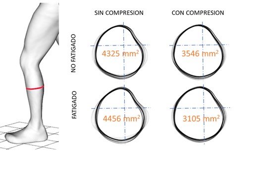
Figure 7. Study of shape variation of the left calf perimeter at 35% under the four conditions studied, and SVI value (in orange)
Finally, a morphological analysis of the calf perimeter contour was performed in relation to the different phases of the running cycle. The results showed a direct correlation between contour configuration and the corresponding biomechanical phase. Specifically, during the stance phase, the calf contour adopts a more compact and circular configuration, suggesting greater tissue tension and a homogeneous redistribution of muscle volume. By contrast, during the aerial phases of the running cycle, the contour elongates significantly along one of its main axes, reflecting muscle relaxation and passive displacement of soft tissue (see Figure 8). This dynamic contour behaviour provides quantitative evidence of movement-induced morphological variability and reinforces the need to consider these deformations in the design of functional compression garments.
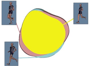
Figure 8. Study of the perimeter shape associated with key instants of the movement
CONCLUSIONS
Soft-tissue displacement can negatively affect sports practice, as it is associated with reduced biomechanical efficiency, increased perceived neuromuscular fatigue and a higher risk of injury. However, its identification and quantification remain a technically complex challenge and, to a large extent, an unresolved issue to date.
With this motivation, IBV has designed an innovative methodology that makes it possible to locate and quantify soft-tissue displacement during the execution of sports movements. The methodology is based on MOVE4D technology, which enables global and direct assessment of the morphological variations associated with such displacement. Furthermore, as a non-intrusive technique, it does not interfere with technical execution or condition the athlete’s natural movement.
The methodology has undergone preliminary validation via a pilot study carried out with a small number of amateur runners. The results have confirmed the technical feasibility of the system and have enabled the identification and definition of the key biomechanical variables required for the spatial and temporal quantification of soft-tissue displacement. The information generated not only enables estimation of displacement magnitude, but also mapping of its anatomical location.
This information can be used to design more effective and comfortable compression garments by defining a compression-needs map for each body segment, optimising both biomechanical effectiveness and perceived user comfort.
ACKNOWLEDGEMENT
This project is supported by the Regional Ministry of Innovation, Industry and Tourism of the Generalitat Valenciana, through IVACE+i, and is funded by the European Union through the FEDER Comunitat Valenciana 2021–2027 Programme (IMDEEA/2024/24).

BIBLIOGRAPHY
- Chadefaux D, Gueguen N, Thouze A, Rao G. 3D propagation of the shock-induced vibrations through the whole lower-limb during running. J Biomech. (2019); 96:109343. https:// doi. org/ 10. 1016/j. jbiom ech. 2019109343.
- Play, M., Giandolini, M., Perrin, T.P., METRA, M., Feasson, L., Rossi, J., Millet, G.Y.: Soft‐Tissue Vibrations and Fatigue During Prolonged Running: Does an Individualized Midsole Hardness Play a Role? Scandinavian Journal of Medicine & Science in Sports. (2024). https://doi.org/10.1111/sms.14672.
- Ehrström, S., Gruet, M., Giandolini, M., Chapuis, S., Morin, J.-B., Vercruyssen, F.: Acute and Delayed Neuromuscular Alterations Induced by Downhill Running in Trained Trail Runners: Beneficial Effects of High-Pressure Compression Garments. Frontiers in Phy-siology. (2018). https://doi.org/10.3389/FPHYS.2018.01627
- Dewolf, A. H., Ivaniski-Mello, A., Peyré-Tartaruga, L.A., Mesquita, R.M.: Relation between soft tissue energy dissipation and leg stiffness in running at different step frequencies. Royal Society Open Science, 11. (2024). https://doi.org/10.1098/rsos.231736
- Chadefaux, D., Berton, E., Rao, G.: How runners deal with the shock induced vibrations propagating through their body. Journal of the Acoustical Society of America. (2017). https://doi.org/10.1121/1.4989079
- Boyer, K.A., Nigg, B.M.: Muscle Tuning During Running: Implications of an Un-tuned Landing The impact force in heel-toe running is an input signal into the body that. (2006).
- Leabeater, A.J., James, L.P., Driller, M.W.: Tight Margins: Compression Garment Use during Exercise and Recovery—A Systematic Review. Textiles. (2022). https://doi.
- Engel, F.A., Stockinger, C., Woll, A., Sperlich, B.: Effects of Compression Garments on Performance and Recovery in Endurance Athletes. (2016). https://doi.org/10.1007/978-3-319-39480-0_2.
- Hong, W.-H., Lo, S.-F., Wu, H.C., Chiu, M.-C.: Effects of compression garment on muscular efficacy, proprioception, and recovery after exercise-induced muscle fatigue onset for people who exercise regularly. PLOS ONE. (2022). https://doi.org/10.1371/journal.pone.0264569.
- Doan, B.K., Kwon, Y.-H., Newton, R.U., Shim, J.K., Popper, E.M., Rogers, R.A., Bolt, L.R., Robertson, M.C., Kraemer, W.J.: Evaluation of a lower-body compression gar-ment. Journal of Sports Sciences. (2003). https://doi.org/10.1080/0264041031000101971.
- Play MC, Trama R, Millet GY, Hautier C, Giandolini M, Rossi J. Soft Tissue Vibrations in Running: A Narrative Review. Sports Med Open. (2022);8(1):131. doi: 10.1186/s40798-022-00524-w. PMID: 36273049; PMCID: PMC9588116.
- Human body 4D Scanning technology. https://www.move4d.net/es/move-4d/
AUTHOR’S AFFILIATION
Instituto de Biomecánica de Valencia
Universitat Politècnica de València
Edificio 9C. Camino de Vera s/n
(46022) Valencia. Spain
* Universidad Europea de Valencia
HOW TO CITE THIS ARTICLE
Author/s: Alfredo Remón Gómez, José Pérez Maletzki*, Alfredo Ballester Fernández, Javier Gámez Payá, Sandra Alemany Mut. (15 December 2025). «MOVE4D: dynamic scanning technology for quantifying the effect of compression garments in runners.» Revista de Biomecánica nº 72. https://www.ibv.org/actualidad/move4d-la-tecnologia-de-escaneado-dinamico-que-cuantifica-el-efecto-de-las-prendas-de-compresion-en-corredores/

The publication of this article is funded by Budget Line S1048, “Technology Centres of the Valencian Community. Targeted support”, of the Valencian Regional Government’s 2025 Budget (IMAMCA/2025/7).