Nowadays, Anterior Cruciate Ligament (ACL) injuries remain a challenge that requires exhaustive functional evaluation to guarantee recovery and prevent future re-injuries. In this context, dynamic body surface scanning technology (MOVE4D) emerges as a valuable and innovative tool in biomechanical analysis. This system allows the quantification of the body’s three-dimensional morphology in motion, offering a unique perspective. The study of dynamic morphology provides significant information on structural variables (such as atrophy or edema) and functional variables (soft tissue deformation). In this context, dynamic morphological analysis enhances the precision of post-ACL assessment and guides more objective decisions regarding functional progression.
INTRODUCCIÓN
Anterior cruciate ligament (ACL) injuries of the knee are currently considered one of the most common pathologies both in sports and in the general population. They frequently occur during movements that involve jumping, pivoting, or sudden changes of direction, whether in sports or work settings, and are often associated with physical exertion, movement on uneven surfaces, or work-related accidents. These injuries typically lead to alterations that affect the biomechanics of the knee and the functional capacity of the lower limb. Therefore, proper assessment and follow-up of this injury are essential not only for returning to sports but also for regaining adequate function in daily or work-related activities. In this regard, it is not sufficient to assess only the integrity of the ligament reconstruction. Dynamic evaluation through movement patterns is essential, as well as monitoring of the musculature to ensure a safe return, with the goal of reducing the risk of re-injury or joint degeneration. There are examples in the literature [1,2] that highlight morphological and physiological changes—such as reductions in strength, cross-sectional area, and volume—as long-term negative effects of ACL injuries. Several studies have shown that, after an ACL injury or reconstruction, gait kinematics may approach a nearly symmetrical pattern between the affected and contralateral limbs, especially in advanced stages of recovery. However, this apparent normalization of joint kinematics does not imply a complete restoration of muscle function [3,4]. In routine clinical practice, follow-up after ACL reconstruction continues to rely mainly on time-based criteria to estimate recovery, complemented by strength and functional tests, as well as instrumental biomechanical assessments. Currently, when assessing and monitoring ACL injuries through motion-capture technologies—3D stereophotogrammetry, IMUs, and markerless systems—information on joint kinematics is obtained, but this is influenced by limitations such as soft tissue artifact, sensor drift/placement, and accuracy that varies depending on the task and the population. On the other hand, in muscle assessment, surface electromyography is sensitive to crosstalk and electrode placement despite existing guidelines for its standardized use. All these factors may influence, to a greater or lesser extent, the reliability and accuracy of these analyses.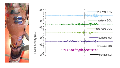
Figura 1. Ejemplo de medición de la actividad muscular con electromiografía.
DESARROLLO
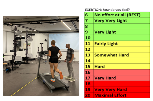
Figura 2. Imagen de una sesión de captura de laboratorio (izquierda) y escala Borg 6-20 para la valoración de la fatiga.
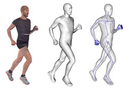
Figura 3. Ejemplo de procesado de un frame: datos capturados (izquierda), avatar procesado (centro) y esqueleto (derecha).
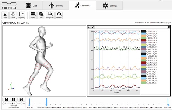
Figura 4. Representación sobre el avatar de los perímetros calculados (iquierda) y gráfica con los valores de los perímetros en la secuencia capturada (derecha).
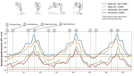
Figura 5. Estudio de la longitud del perímetro del gemelo izquierdo 35% en las 4 condiciones estudiadas.
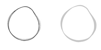
Figura 6. Perímetros de la secuencia alineados (izquierda). Área encerrada por los perímetros o SVI (derecha).
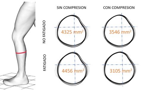
Figura 7. Estudio de la variación de forma del perímetro del gemelo izquierdo 35% en las 4 condiciones estudiadas y valor del SVI (en naranja)
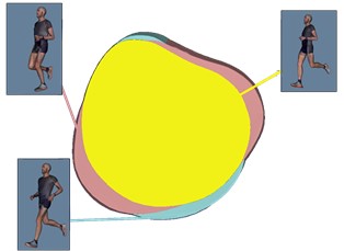
Figura 8. Estudio de la forma del perímetro asociado a los momentos clave del movimiento.
CONCLUSIONES
AGRADECIMIENTOS

BIBLIOGRAFIA
- Chadefaux D, Gueguen N, Thouze A, Rao G. 3D propagation of the shock-induced vibrations through the whole lower-limb during running. J Biomech. (2019); 96:109343. https:// doi. org/ 10. 1016/j. jbiom ech. 2019109343.
- Play, M., Giandolini, M., Perrin, T.P., METRA, M., Feasson, L., Rossi, J., Millet, G.Y.: Soft‐Tissue Vibrations and Fatigue During Prolonged Running: Does an Individualized Midsole Hardness Play a Role? Scandinavian Journal of Medicine & Science in Sports. (2024). https://doi.org/10.1111/sms.14672.
- Ehrström, S., Gruet, M., Giandolini, M., Chapuis, S., Morin, J.-B., Vercruyssen, F.: Acute and Delayed Neuromuscular Alterations Induced by Downhill Running in Trained Trail Runners: Beneficial Effects of High-Pressure Compression Garments. Frontiers in Phy-siology. (2018). https://doi.org/10.3389/FPHYS.2018.01627
- Dewolf, A. H., Ivaniski-Mello, A., Peyré-Tartaruga, L.A., Mesquita, R.M.: Relation between soft tissue energy dissipation and leg stiffness in running at different step frequencies. Royal Society Open Science, 11. (2024). https://doi.org/10.1098/rsos.231736
- Chadefaux, D., Berton, E., Rao, G.: How runners deal with the shock induced vibrations propagating through their body. Journal of the Acoustical Society of America. (2017). https://doi.org/10.1121/1.4989079
- Boyer, K.A., Nigg, B.M.: Muscle Tuning During Running: Implications of an Un-tuned Landing The impact force in heel-toe running is an input signal into the body that. (2006).
- Leabeater, A.J., James, L.P., Driller, M.W.: Tight Margins: Compression Garment Use during Exercise and Recovery—A Systematic Review. Textiles. (2022). https://doi.
- Engel, F.A., Stockinger, C., Woll, A., Sperlich, B.: Effects of Compression Garments on Performance and Recovery in Endurance Athletes. (2016). https://doi.org/10.1007/978-3-319-39480-0_2.
- Hong, W.-H., Lo, S.-F., Wu, H.C., Chiu, M.-C.: Effects of compression garment on muscular efficacy, proprioception, and recovery after exercise-induced muscle fatigue onset for people who exercise regularly. PLOS ONE. (2022). https://doi.org/10.1371/journal.pone.0264569.
- Doan, B.K., Kwon, Y.-H., Newton, R.U., Shim, J.K., Popper, E.M., Rogers, R.A., Bolt, L.R., Robertson, M.C., Kraemer, W.J.: Evaluation of a lower-body compression gar-ment. Journal of Sports Sciences. (2003). https://doi.org/10.1080/0264041031000101971.
- Play MC, Trama R, Millet GY, Hautier C, Giandolini M, Rossi J. Soft Tissue Vibrations in Running: A Narrative Review. Sports Med Open. (2022);8(1):131. doi: 10.1186/s40798-022-00524-w. PMID: 36273049; PMCID: PMC9588116.
- Human body 4D Scanning technology. https://www.move4d.net/es/move-4d/
AFILIACIÓN DE LOS AUTORES
Instituto de Biomecánica de Valencia Universitat Politècnica de València Edificio 9C. Camino de Vera s/n (46022) Valencia. Spain * Universidad Europea de ValenciaCÓMO CITAR ESTE ARTÍCULO
Autor/es: Alfredo Remón Gómez, José Pérez Maletzki*, Alfredo Ballester Fernández, Javier Gámez Payá, Sandra Alemany Mut. (15 de Diciembre de 2025). «MOVE4D: la tecnología de escaneado dinámico que cuantifica el efecto de las prendas de compresión en corredores. Revista de Biomecánica nº 72. https://www.ibv.org/actualidad/move4d-la-tecnologia-de-escaneado-dinamico-que-cuantifica-el-efecto-de-las-prendas-de-compresion-en-corredores/ The publication of this article is funded by Budget Line S1048, “Technology Centres of the Valencian Community. Targeted support”, of the Valencian Regional Government’s 2025 Budget (IMAMCA/2025/7).